
Abstract
LLNL has recently licensed its hand-held advanced nucleic-acid analyzer, the HANAA, for commercial development. This (as is characteristic of other miniaturized, portable diagnostic instruments being developed around the world) typically still requires manual sample handling and preparation by skilled operators. Dielectrophoretic and acoustic manipulation of fluids and particles are techniques that have shown good promise to reduce the demands on manual sample preparation both for miniature and for table-top instrumentation.
INTRODUCTION
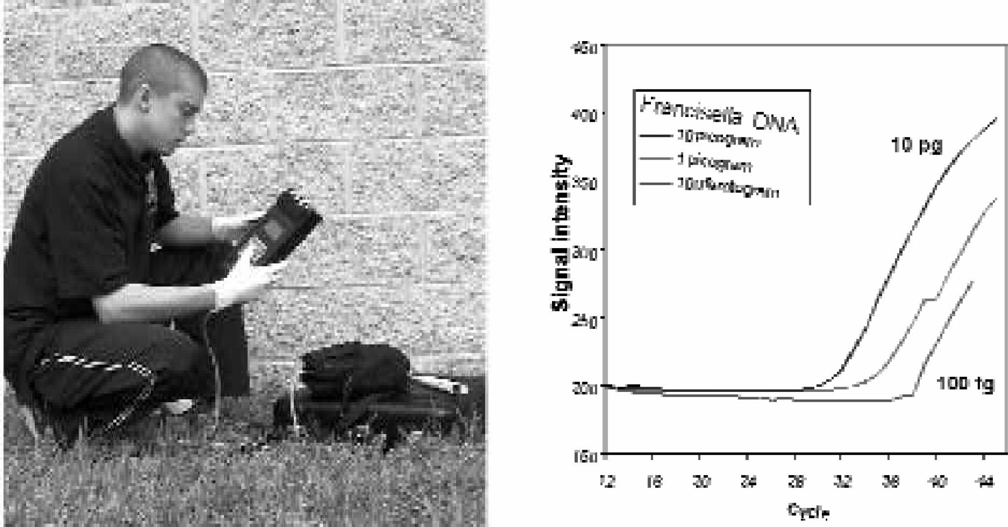
The world relies on immunoassays and nucleic-acid assays for a wide range of biomedical applications. Many advances have been made in the instrumentation that performs such assays, including detection technology, but the up-front effort in sample preparation remains an impediment. Figure 1 shows the hand-held advanced nucleic-acid analyzer, the HANAA, and data collected with it in a laboratory setting in January, 2000. The HANAA is a four-chamber, battery-powered instrument that performs realtime polymerase-chain-reaction assays. Figure 2 shows the HANAA in field use, along with data collected at the U.S. Army Edgewood Chemical Biological Center, Soldier Biological and Chemical Command, at Aberdeen Proving Grounds 1 . Note that the technically-skilled person who is using the HANAA, has a micropipetter nearby. Manual sample handling and manual sample preparation are particular problems in a field setting, where the sample being processed is truly an “environmental” sample – contaminated with both natural and artificial debris such as dirt or soot or other combustion products and by-products. While it is inviting to imagine an integrated microfluidics system that will collect, handle, and prepare such samples as well as perform the assays, themselves, the reality is that clogging of microchannels can thwart this approach, and contamination and non-specific binding can interfere with the assays.
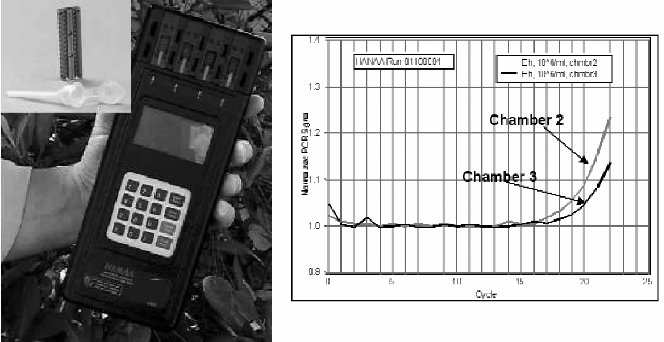
Left side: Photograph of the battery-powered, hand-held, realtime PCR instrument, HANAA (hand-held, advanced, nucleic-acid analyzer). The insert shows a photograph of the HANAA's silicon thermal cycling chamber and polypropylene sample tube. This chamber has two identical halves that are not bonded together, but rather are spring-loaded to press against the sides of the plastic sample tube. The etching that penetrates entirely through each half permits cooling air to pass more uniformly around the sample tube when the fan is operating and also reduces the overall thermal mass of the system. There are two windows, providing optical access to the sample tube, shown below the chamber. The polypropylene sample tube contains up to 25 μL of solution. The dimensions of the silicon chamber are 2 mm thick, 6.5 mm wide, and 23 mm long.
Left side: Photograph of a HANAA in use at the U.S. Army Edgewood Chemical Biological Center (ECBC), Soldier Biological and Chemical Command, at Aberdeen Proving Grounds.
Clogging of microchannels by debris looms as a serious problem when the dimensions of channels that transport the sample become much less than one mm – even though such microfluidic channels play important roles in the miniaturization of the instruments that perform bioassays. Figure 3 shows a recent autonomous pathogen detection system (APDS) designed and built for the U. S. Department of Energy Chem-Bio National Security Program at LLNL, along with 4-plex immunoassay data it collected during a 24-hour run. The APDS continuously collects air samples, performs sample preparation for immunoassay, and runs the multiplex immunoassay on the Luminex, Inc., flow cytometer. This instrument stands roughly 1.5 meters high and uses millimeter diameter tubing for sample handling. Although we desire to miniaturize this system, we wish to minimize any decrease in its performance, including resistance to clogging.
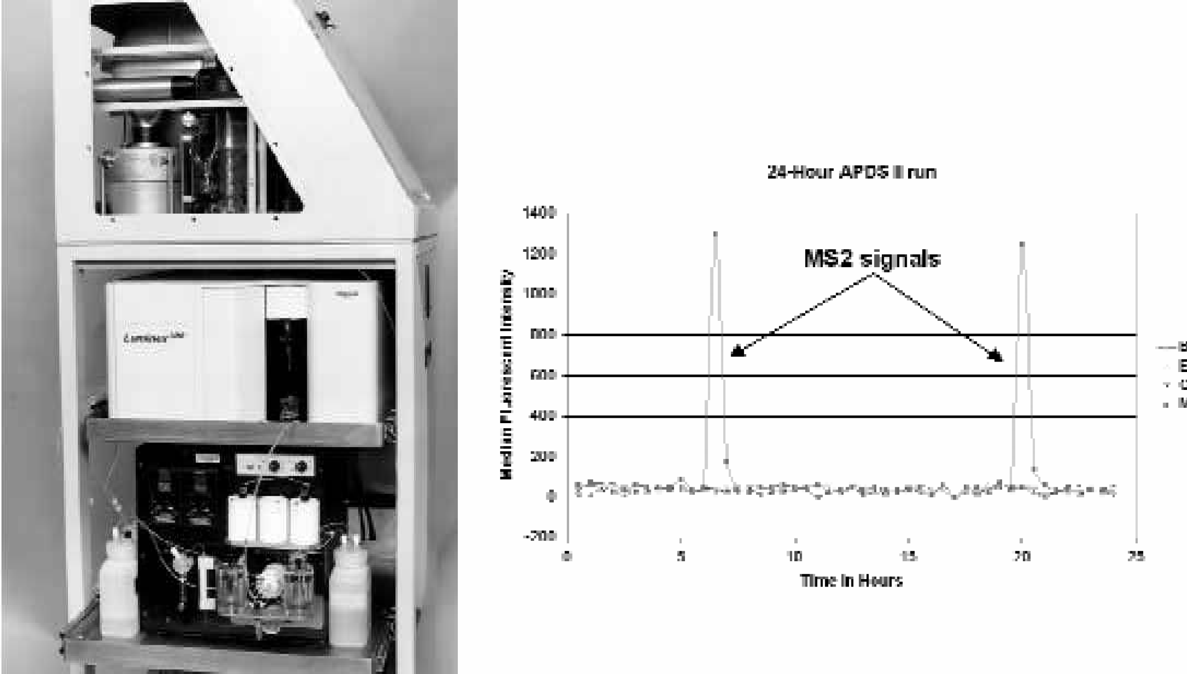
Left side: Photograph of autonomous pathogen detection system (APDS). The aerosol collector is visible on the top shelf, with the flow cytometer placed on the middle shelf and the fluidics on the bottom shelf.
Those of us who have had to analyze hundreds of unknowns through any system with a critical orifice that is less than 100 μm have faced the problem of clogging. As an example, a team from LLNL participated in the Department of Defense's Joint Field Trials III, in 1996. One of the instruments that we operated was a flow cytometer that had a critical, round orifice with a diameter of 50 μm. We used this flow cytometer to detect, identify, and quantify every unknown with either bacterial spores or live bacteria over the entire concentration range of unknowns (10 3 /ml through 10 6 /ml). To reduce the danger of clogging our instrument, we hand-filtered every unknown through a disposable, single-use porous filter that nominally removed particles larger than 20 μm. Almost every day of the 10 days of the trials, we still experienced clogging of the critical orifice. When we examined the clogged orifice under a microscope, the reason for this problem became clear – some debris was not round. The debris that was long and thin could easily have an end-view diameter that was less than 20 μm, while sporting a length of more than 50 μm. Some of these microscopic “logs” passed through the filter and created a logjam in the fluidics. For such circumstances and many others, it is desirable to be able to separate the biological materials in an unknown solution from the debris that may also be present.
Both immunoassays and nucleic-acid-based assays are amenable to microliter sample volumes and, therefore, are suitable for miniaturization. However, corresponding sample preparation techniques must accompany this miniaturization. With a miniature system it is particularly desirable to avoid the use of physical filters/filter paper and/or centrifugation for sample preparation. Dielectrophoresis and acoustic manipulation of fluids and particles are examples of techniques that promise to become integral parts of miniaturized instruments that include sample handling and sample preparation as well as bioassays.
Acoustic Manipulation of Particles
We are able to combine high functionality with an inexpensive, replaceable sample chamber when we remotely couple acoustic energy into a glass or plastic chamber. 2 Particles are manipulated by acoustic radiation pressure. Radiation pressure generated in the presence of a standing wave induces a pressure field that forces particles to collect in the nodes or anti-nodes of the standing wave. The acoustic force is a function of particle size, density, and acoustic energy; hence, acoustic radiation pressure can be used to transport particles as well as to differentiate particles based on their size and material attributes. These functions have been demonstrated previously in macro- and MEMS-based devices. 3 –6 We have demonstrated a non-contact method of concentrating particles in a microfluidic chamber employing acoustic radiation pressure. This, under the typical conditions of laminar flow in microchannels, enriches the particles in solution away from the dissolved components. Although it is speculation at this point, it may also be beneficial to expose an environmental sample to acoustic energy in order to “de-clump” important biological targets such as bacteria or viruses and to dislodge them from adhering (and interfering) debris.
We have fabricated and tested prototype devices for acoustic-based particle manipulation that indicate preferential geometry for the sample-handling function of concentrating. Typical devices consist of a single half-wavelength width fluid channel fabricated from laser-cut acrylic sheets or glass. Bulk PZT-4 piezoelectric transducers were used as the acoustic source. One prototype device is shown in Figure 4. Piezoelectric transducers had a half-wavelength resonance thickness and exhibited a primary resonance peak at 365 kHz. A flow cell package has also been designed that integrates reusable liquid sample interconnects [see reference 2], electrical contacts, and a removable sample chamber.
Photograph of a microfluidic-trapping chamber. The bonded glass package is 1 cm X 2 cm X 3 mm.
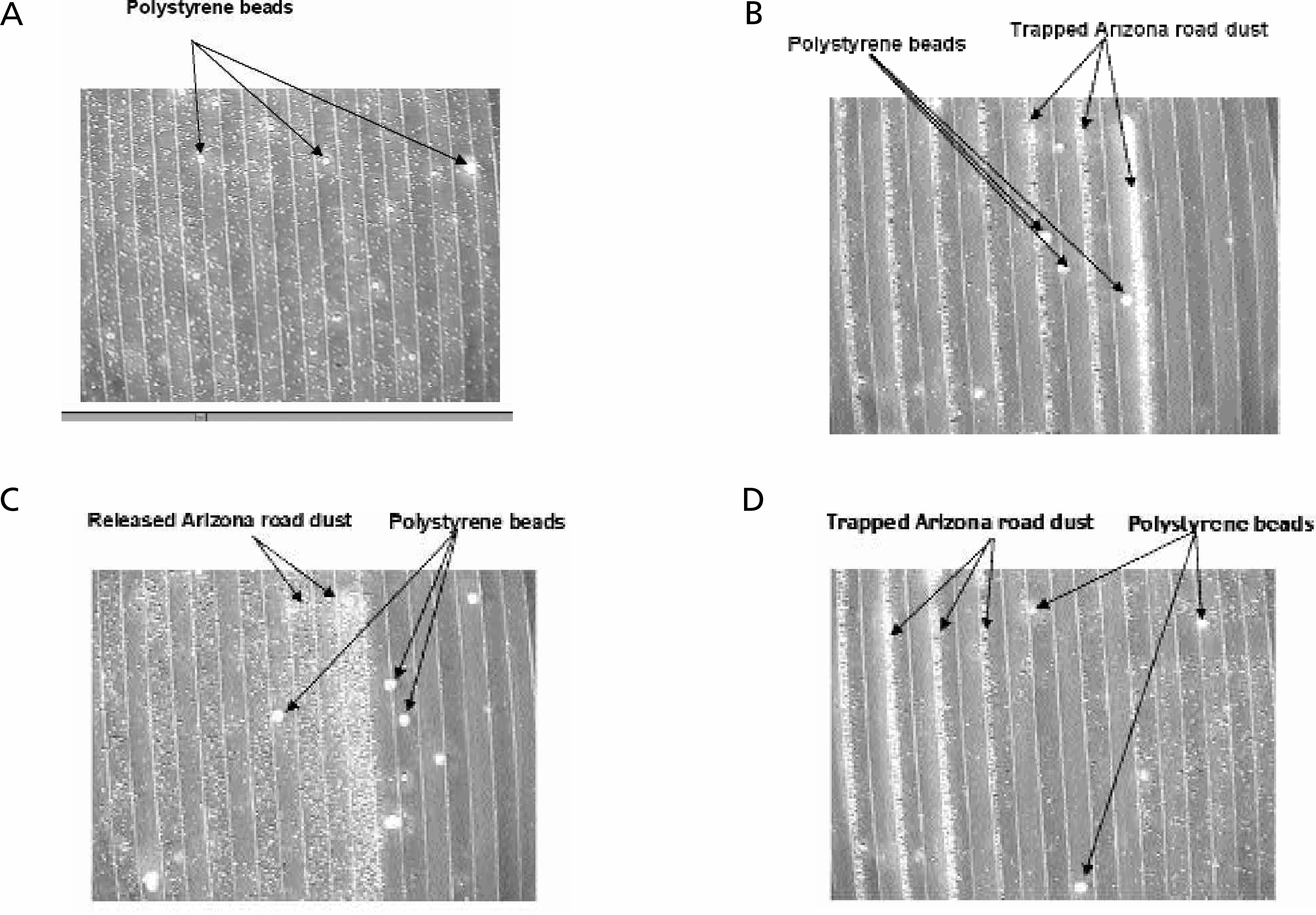
We have been able to concentrate 10 μm polystyrene beads at the center of the fluid channel, an expected pressure node of the standing wave. We have also observed separation of particles based on size. The larger beads collect at the central node of the channel, while the smaller, 3 μm beads remain dispersed in solution. (see Figure 5).
Left side: Photomicrograph of the 10 μm beads in the fluidic channel from Figure 4, before the piezoelectric elements are activated.
Since the flow rates and dimensions are small enough to ensure laminar flow, it would be straightforward to pass such a separated and concentrated suspension through a dual-orifice exit, where the beads would be separated from the bulk of the solution. This would constitute a form of a wash step in sample preparation.
DIELECTROPHORESIS
Dielectrophoresis is a technique that exploits the different polarizabilities of materials in order to manipulate them. 7 All materials have some polarizability, and, under the influence of an heterogenous electric field, this can provide differential forces between components of a fluidic mixture. With this technique, blood cells and other particles in solution have successfully been manipulated and separated. 8 –13 Typically, dielectrophoresis relies on an alternating voltage, so that the permanent charge on a molecule or particle does not dominate the control of its movements.
The time-averaged dielectrophoretic force is a function of the gradient in the electric field, and is not a function of the applied voltage. For this reason, dielectrophoresis in aqueous solutions is suitable for miniature aqueous fluidic systems, and is not well suited for macroscopic aqueous fluidic systems. (To achieve high field gradients in macroscopic systems it would be necessary to apply large voltages, which cause electrolysis of water, interfering with most separation and manipulation procedures). Figure 6 shows a photograph of an integrated package that includes a micropump, fluidic channels, and dielectrophoresis electrodes. Using gold dielectrophoresis electrodes (30 μm in width and 30 μm in separation) and relatively low-frequency alternating voltage (under 1 kHz, less than 2 Vrms), we have trapped bacterial spores. 14
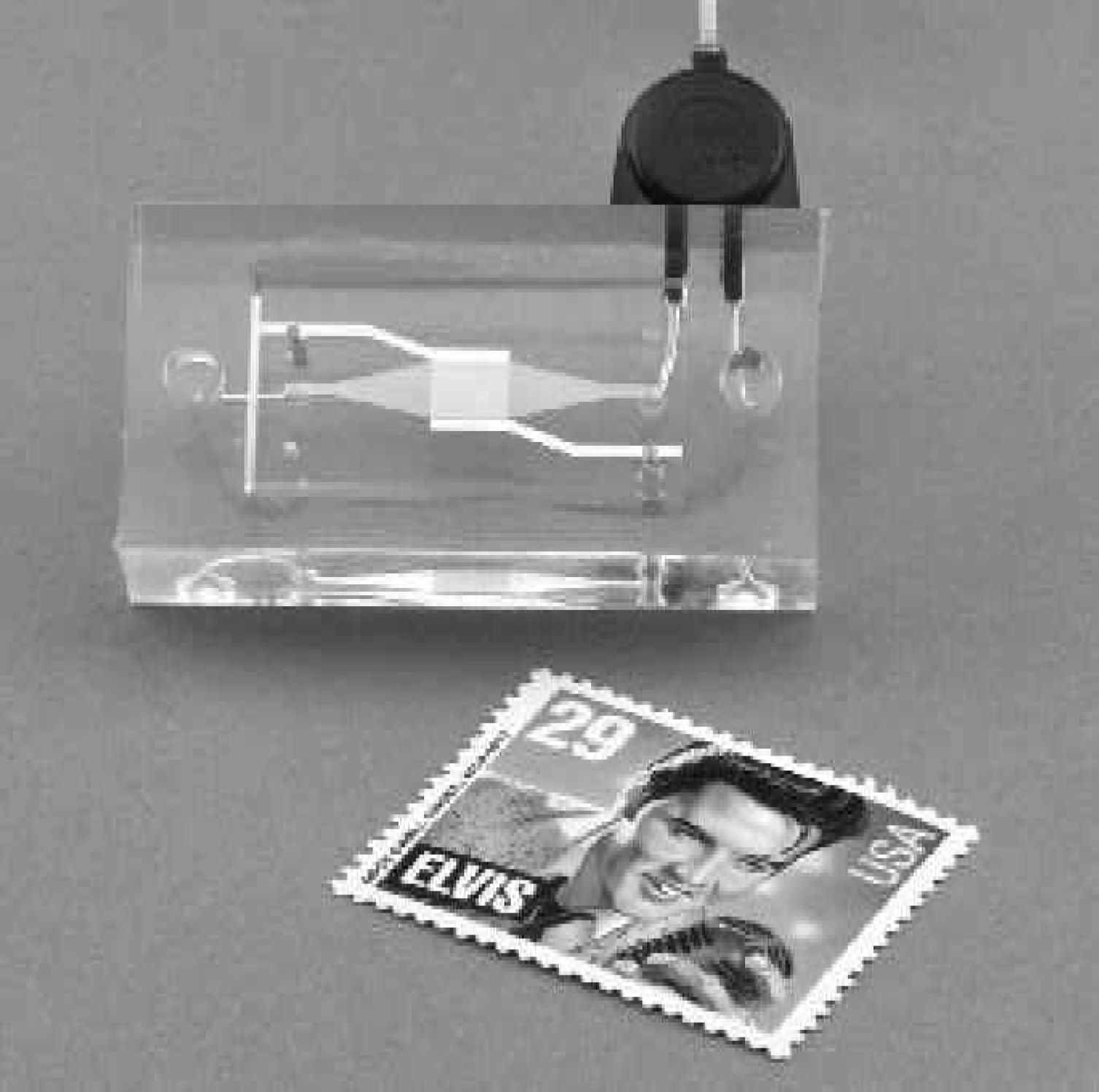
Photograph of a integrated fluidics package, with micropump, flow channels, and dielectrophoresis electrodes. The US postage stamp is included to show the size of the package.
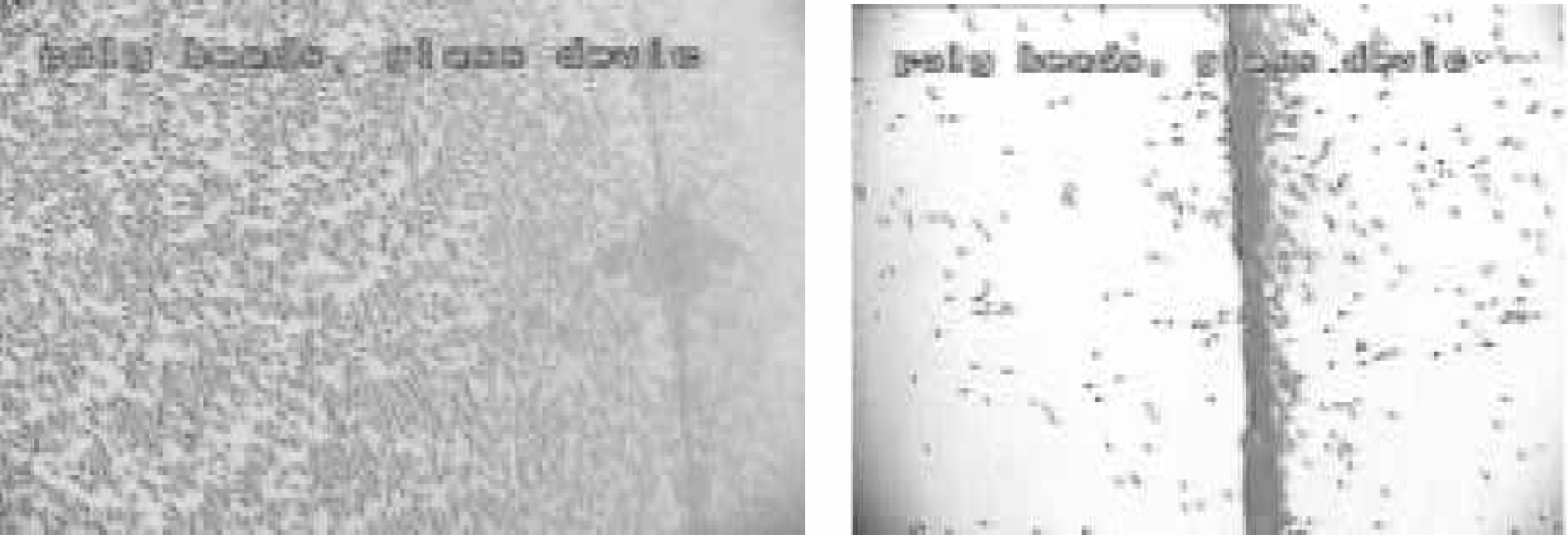
Using a higher-frequency alternating electric field, we were able to take a mixture of Arizona road dust and 3.4 μm-diameter polystyrene microspheres and process this mixture through a flow cell with dielectrophoresis electrodes – the dielectrophoretic effect removed the Arizona road dust preferentially from the solution. For the handling/preparation of environmentally contaminated samples, this is very important. Figure 7 displays a timed series of micrographs that were taken in the flowing system as the dielectrophoresis electrodes are alternatively “off” or “on”.
A sequence of photomicrographs of a flowing solution of Arizona road dust (ARD) and 3.4-μm polystyrene beads through the dielectrophoresis chamber. The ARD, present in much higher concentration, is typically about 1 μm in dimension. Flow direction is from right to left. Figure 7a shows the distribution of particles before the dielectrophoresis is activated. Figure 7b shows the distribution of particles when the dielectrophoresis is activated. Note the extensive deposits of ARD on a few dielectrophoresis electrodes. Figure 7c shows the same chamber, 10 seconds after the photomicrograph in Figure 7b was taken, when the dielectrophoresis is, again, turned off. The ARD has been released back into the flowing solution. Figure 7d shows the same chamber, 10 seconds after the photomicrograph in Figure 7c was taken. The ARD is re-captured, but on electrodes that are farther downstream.
CONCLUSIONS
Although research in portable instrumentation has produced miniature versions that match or even exceed the performance of larger, more power-hungry equipment, the collection, handling, and preparation of environmentally-contaminated samples continues to be performed manually in most cases. Nonetheless, new techniques such as dielectrophoresis and acoustic manipulation of particles are offering to increase the amount of work performed within an integrated system and to decrease the level of effort and training that is demanded of the operator. The inclusion of such techniques into an autonomous systems is on the horizon.
ACKNOWLEDGEMENTS
The author gratefully acknowledges the many contributions to this subject by William Benett, Kerry Bettencourt, John Chang, Julie Hamilton, Dawn Hilken, Ron Koopman, Peter Krulevitch, Fred Milanovich, Jim Richards, Klint Rose, Paul Stratton, Lisa Tarte, Kodumudi Venkateswaran, Steven Visuri, Amy Wang, and Tom Wilson. This work was performed under the auspices of the U.S. Department of Energy by University of California Lawrence Livermore National Laboratory under contract No. W- 7405-Eng-48.