
Abstract
Thalassemia and malaria are present at high frequency in tropic and subtropic regions. Impaired growth of Plasmodium falciparum has been demonstrated in vitro in both α-and β- thalasemic erythrocytes. In this study, we investigated the effects of thalassemic sera on Plasmodium falciparum developement including 5 α-thalasemia1/α-thalasemia2 (HbH) sera, 4 a-thalasemia1/ hemoglobin Constant Springs (HbH/HbCS) sera and 7 β-thalassemia/ HbE sera compared to 7 normal sera. Study on malarial growth in medium containing 10% various sera for 4 days revealed that HbH and b-thalasemia/ HbE sera was significantly inhibited (p<0.05, by Mann-Whitney Utest) the parasite growth comparing to normal serum. Mean percentages of the parasites grown in HbH (2.24+0.75%) and β-thalassemia/ HbE (1.69+0.97%) sera were about 2 times less than that in normal serum (4.55+2.40%). The mean parasitemia cultured in HbH/HbCS serum (2.38+1.54%) was also lower than that in normal serum but it was non-significantly different. Investigation on stage changing of the parasite among the starting, 1st, 2nd, 3rd and 4th day showed that all thalassemic sera retarded the parasite maturation from stage to stage. Decrease in percent ring stage productionwas observed in culture with HbH/HbCS and β-thalassemia/HbE sera. This might be resulted from reduction of merozoite population in mature schizont or failure of re-invasion of the merozoite. Therefore, not only cellular factors but also serum factors of thalassemic patients inhibited P. falciparum development which may be related to the protective mechanism of thalassemic patients from P. falciparum infection
Keywords
INTRODUCTION
Haldaneís malaria hypothesis explains high frequency of thalassemia in tropical and subtropical regions is due to natural selection by malaria(1). Several investigations showed that erythrocytes of thalassemia and hemoglobinopathies were less susceptible to Plasmodium falciparum infection than those of normal hemoglobin. Impaired growth of P.falciparum has been demonstrated in vitro in either α- and β-thalassemic erythrocytes or in erythrocytes containing hemoglobin (Hb) CS, Hb S, Hb C and Hb F(2 –8). Protective mechanisms of these variant erythrocytes may occur at the level of invasion of erythrocytes and intraerythrocytic development(2 –9). Host immune response, reologic and other extraerythrocytic determinants may also involve(2,3,8,10). Only few studies have explored the effect of the variant sera on P.falciparum growth(11,12). Kaminsky et al. reported that serum from a heterozygote β-thalassemic patient could inhibit P.falciparum growth in vitro when the parasite was cultured only in the β-thalassemic erythrocytes. That inhibitory effect was abolished in normal erythrocytes(11). Contradictorily, Thanomsub et al. demonstrated suppressive effects of thalassemic sera (both α- and β-) on P.falciparum life-cycle in both variant and normal erythrocytes. They also demonstrated that sera from classical hemoglobin H patients showed most effective in suppressing parasite growth(12). Here, we clearly show the inhibitory effect of thalassemic sera on growth retardation and development of P.falciparum in vitro. However the most inhibitory effect was demonstrated in β-thalasemia/HbE serum.
MATERIALS AND METHODS
RESULTS
After culturing P.falciparum in the media containing various tested sera for 4 days, the mean percent parasitemia of the media with thalassemic sera were about 2 times lower than those with normal serum. The mean percentages of parasites cultured in H-serum (2.24±0.75%) and β/E-serum (1.69±0.97%) were significantly lower than those of normal serum (4.55±2.40%, p<0.05), but was non-significant in H/CS-serum (2.38±1.54%), (Figure 1). The mean of percent parasitemia in the media with normal serum was higher than that with thalassemic sera on the 3rd and 4th days of culture (Figure 2 and table 1). Figure 3 and table 1 showed the mean percentages of ring stage (denoted as R) and trophozoite/schizont stages (denoted as T/S) on each cultured day. Starting from 1% ring stage, the numbers of ring parasites in the next life cycle (the 3rd day) were lower when cultured in H/CS- and β/E-sera than in normal serum (Figure 3B and 3C). This indicated the decrease in merozoite production in mature schizont or failure of re-invasion of the merozoite. Although the pattern of percent ring cultured in H-serum did not clearly lower than that in normal serum, but the percentages of trophozoite/schizont stages in H-serum as well as in H/CS- and β/E-sera were lower than that in normal serum since the 1st day of culture. This indicated parasite growth retardation in culture with α-thalassemic sera.
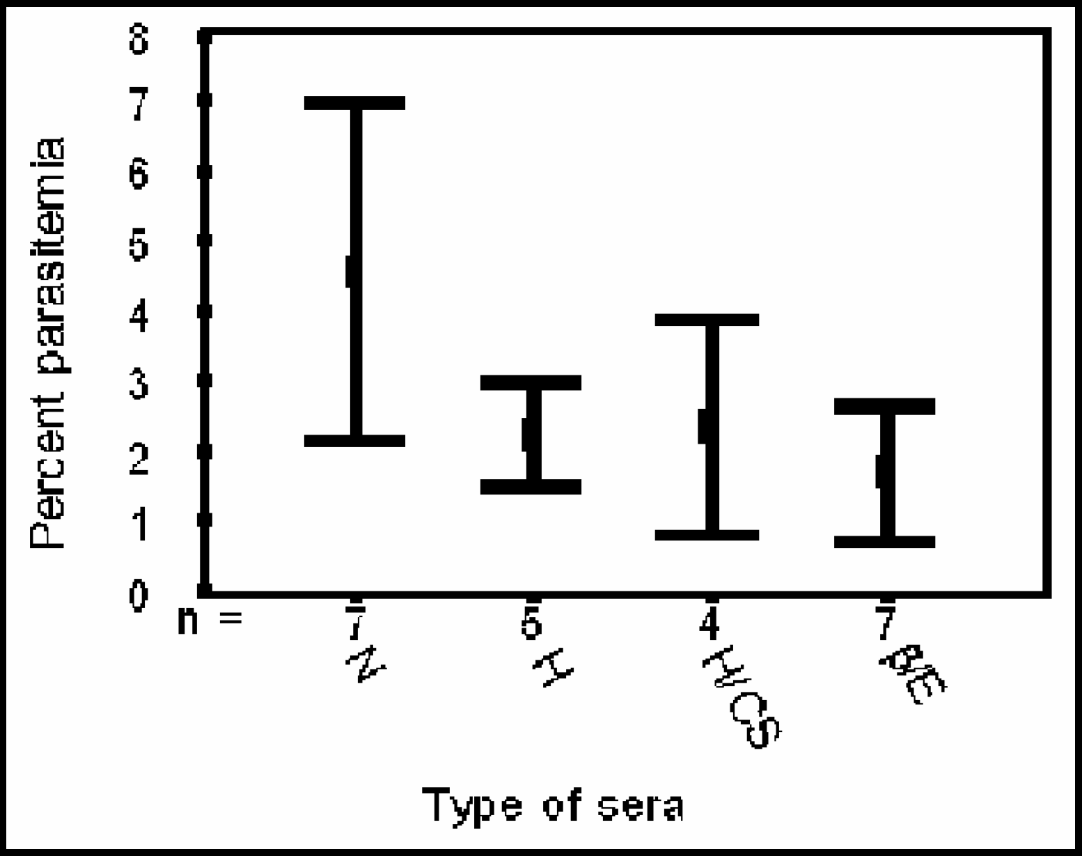
Mean + S.D. of %parasitemia on the 4th day of culture with normal(N) and thalassemic sera (H=HbH, H/CS=HbH/HbCS, and β/E=β-thalassemia /HbE). The mean percent parasitemia culture with thalassemic sera were 2–3 times lower that with normal serum.
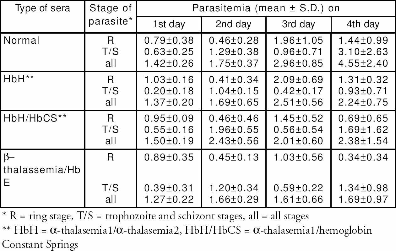
R = ring stage, T/S = trophozoite and schizont stages, all = all stages
HbH = α-thalasemia1/α-thalasemia2, HbH/HbCS = α-thalasemia1/hemoglobin
Constant Springs
DISCUSSION
Innate resistance of thalassemic erythrocytes against plasmodium infection is generally reported(2 –12). In this paper, the effects of thalassemic serum on P. falciparum -cultured in normal erythrocytes were investigated. Interestingly, thalassemic sera significantly inhibit malarial growth at the 2nd intraerythrocytic life cycle or about 4 days of culture (Figure 1). Impaired growth was obviously detected on the 3rd day of culture (Figure 2). These findings are consistent with previous report(12). However, our data revealed contradictory results that the ability of β/E-serum to suppress percent parasitemia was higher than α-thalassemic serum (Figures 1 and 2). Study on malarial stage changing (Figure 3) indicated that ring stage parasites cultured in all thalassemic sera changed to mature stages slower than those of normal serum. In addition, numbers of ring stage in the new parasite life cycle (the 3rd day, Figure 3B and 3C) in thalassemic sera were about 25–50% lower than in normal serum. This indicated that other than thalassemic erythrocytes, thalassemic sera also retarded the parasite development. Thalassemia is one of the diseases associated with iron overload. Excess iron in serum can promote peroxidative damage to cell and organelle membranes(15). In addition depletion of the serum anti-oxidants was also observed(16). Therefore normal erythrocytes cultured in medium with thalassemic serum may be affected by iron induced peroxidative damage. The damaged erythrocytes can be resulted in abortive invasion of the new merozoites, failure to release new merozoites or premature disruption of the sch-izont infected erythrocytes(17). This can lead to decrease in ring-stage numbers in the following life-cycle. Membrane alteration may not only affect the parasite invasion but also affect the intracellular development by altering the transportation of various necessary nutrients(18). Other serum factors such as antibody to neoantigen, complement mediated cell lysis, tumor necrosis factor, etc. can also inhibit malarial development(2,3,18 ).

Mean of %parasitemia on each day of culture with normal(N) and thalassemic sera (H=HbH, H/CS=HbH/HbCS, and β/E= β-thalassemia/HbE). The same numbers of samples in each group as
